Wrist Joint Type Explained: Radiocarpal and Midcarpal
Wrist joint type explained: the radiocarpal and midcarpal joints are condyloid (ellipsoid), how they move, and what this means clinically.

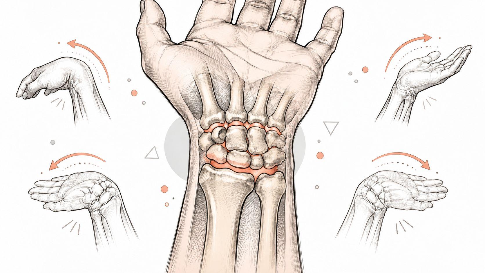
The radiocarpal and midcarpal joints are both condyloid (ellipsoid) joints, which gives the wrist 2 primary degrees of freedom, flexion-extension and radial-ulnar deviation, with no axial rotation of its own. The carpus supplies the machinery: 8 bones in 2 rows, sliding and tilting in a way that no single hinge could manage.
That second number is what makes the standard answer incomplete. Students picture one joint when they hear "wrist," and the anatomy has no single articulation that deserves the name on its own. What sits at the end of the forearm is a family of synovial joints, packed into a small space, sharing ligaments, sharing load, and taking turns producing what looks like one movement. Get the family sorted by type, articulation and direction of movement, and the term "wrist joint type" stops being a phrase to memorise and becomes a description you can reason from.
The confusion that follows people into exams isn't a memory problem. It's a naming problem. Once you know which joint a question is pointing at, the classification follows almost automatically.
Wrist Joint Type at a Glance
The phrase hides two different questions. Are you asking about the radiocarpal joint on its own, or about the functional wrist complex that includes the midcarpal layer? The TeachMeAnatomy wrist joint guide (opens in a new tab) makes the distinction explicit, and it matters because the narrow reading and the broad reading produce different answers to the same exam wording.
In the narrow sense, the radiocarpal joint is a synovial ellipsoid (condyloid) joint, and its main movements are flexion, extension, radial deviation and ulnar deviation. In the broad sense, the wrist is a working group in which the midcarpal joint contributes substantially through intercarpal gliding rather than through a clean hinge action. The two readings don't contradict each other. They describe different scales of the same structure.
Here's the lookup table worth keeping beside your notes.
Reading the table without memorising it
Notice what the first two rows share. Same classification, same pair of movement planes, different contact surfaces. That's the structural reason the wrist can perform circumduction without being a ball-and-socket joint: two planes of tilt, added together, trace a cone.
Now notice what the last two rows don't share with the first two. The thumb carpometacarpal joint sits in the same hand region and gets called part of the wrist in casual conversation, yet it's a saddle joint with opposition as its signature movement. And the distal radioulnar joint is a pivot joint that turns the palm, which is forearm motion, not wrist motion.
Practical rule: if the movement turns the palm or brings the thumb to the little finger, the radiocarpal joint isn't responsible, no matter how close the joint sits to the wrist crease.
That single rule resolves a large share of the marking errors in first-year anatomy answers, and it costs one line to remember.
Why the Wrist Moves Like an Ellipsoid Joint and What Its Bones Do
Picture an egg resting in a shallow cup. The rounded end can tilt forward, back, left and right, but it can't spin on its own axis the way a screw turns. That's the geometry of an ellipsoid joint, and it's the fastest way to feel the difference between the wrist and the shoulder.
The Radiopaedia entry on the radiocarpal joint (opens in a new tab) describes the arrangement precisely. The distal radius and the triangular fibrocartilage articular disc form a proximal concavity, and that cup receives the convex proximal surfaces of the scaphoid, lunate and triquetrum. Two primary rotational degrees of freedom follow from the shape: flexion-extension and radial-ulnar deviation. No third axis appears, which is exactly what the socket geometry forbids.
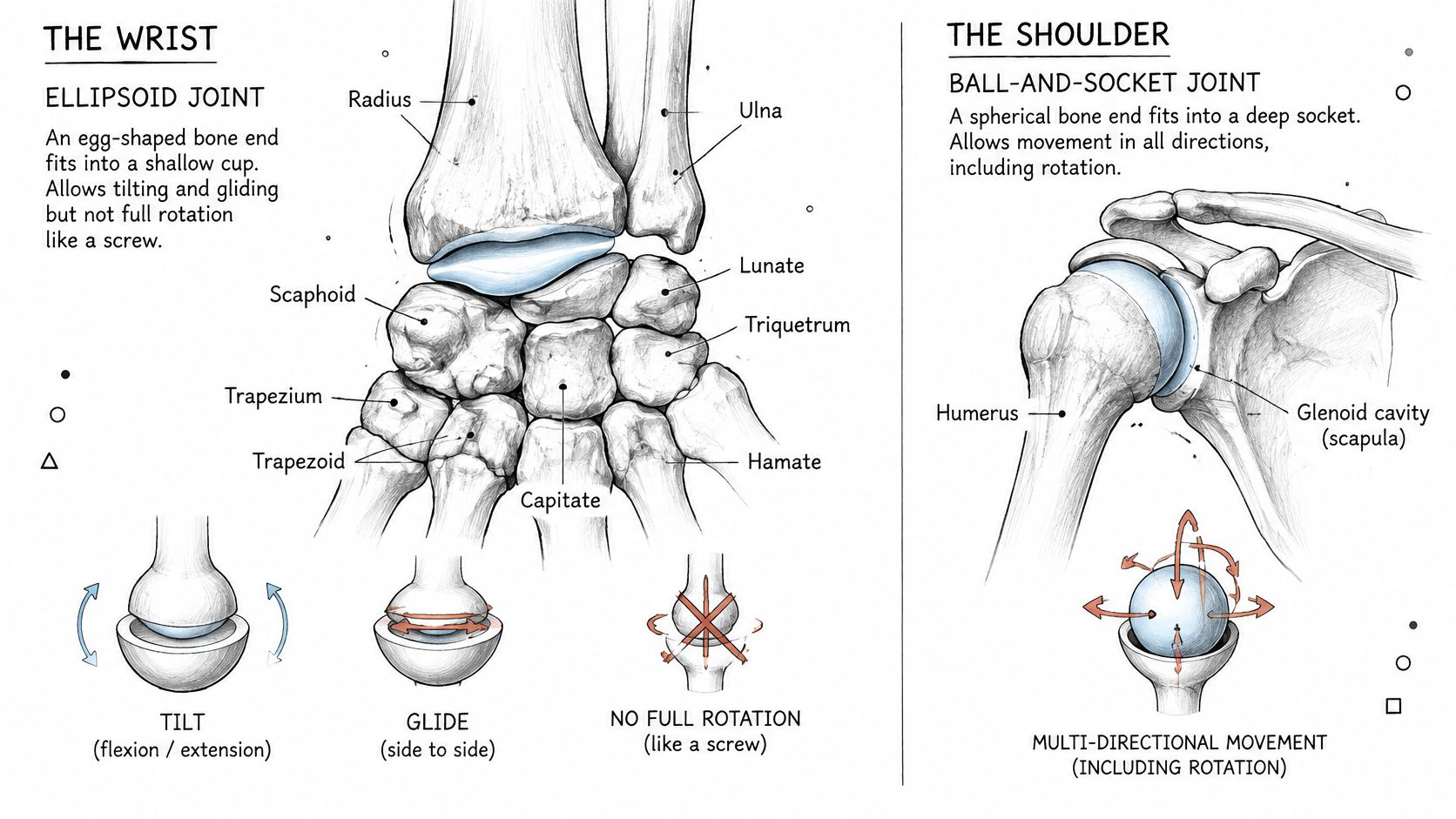
Most texts treat condyloid and ellipsoid as near-synonyms, so a question using either word is asking for the same answer. A ball-and-socket joint adds rotation around the long axis. The wrist doesn't. That's the whole contrast in one line.
The eight bones and the two layers
The anatomy reference on wrist joint structure (opens in a new tab) lays out the carpus as 8 bones in 2 rows. Learn them as four and four, not as a list of eight.
- Proximal row: scaphoid (boat-shaped, at the base of the thumb), lunate (moon-shaped, the bone the radius presses against), triquetrum, and pisiform, a small pea-shaped bone resting on the triquetrum outside the main radiocarpal contact.
- Distal row: trapezium (under the thumb metacarpal), trapezoid, capitate (the largest carpal bone, sitting centrally), and hamate, recognisable by its hook.
Two joint layers sit above and between these rows. The radiocarpal joint sits between the radius and disc above and the proximal row below. The midcarpal joint sits between the two rows, and that's the layer readers forget when they sketch a wrist from memory.
Watch the video on YouTube (opens in a new tab)
A hands-on check you can do now
Find the hollow at the base of the thumb, just distal to the wrist crease, and move your hand toward the thumb side. The bone that surfaces under your fingertip is the scaphoid. It's the bone a clinician checks first after a fall onto an outstretched hand, partly because the blood supply to its proximal portion is limited, which is why a scaphoid injury can heal slowly.
Ligaments bind the rows into a unit with controlled gliding rather than loose stacking. That binding is what converts a pile of small bones into something that behaves like a joint. For a textbook treatment of how these articulations connect to the rest of the skeleton, the Principles of Anatomy and Physiology study document is a useful companion.
Treating the entire wrist as a simple hinge is anatomically inaccurate, and the next section shows why the numbers back that up.
Thumb and Forearm Joints Next to the Wrist
Two joints borrow the wrist's reputation without doing its work.
The distal radioulnar joint is a pivot (trochoid) joint between the distal radius and the head of the ulna. Its movement is pronation and supination, which turns the palm up or down. When you turn a key in a lock, the radius rotates around the ulna; the carpus travels with it but doesn't drive the motion. Calling that wrist rotation is the most common slip in the whole topic.
The thumb carpometacarpal joint is a saddle joint between the trapezium and the first metacarpal. Saddle surfaces are concave in one direction and convex in the other, meeting at right angles, which allows opposition, abduction and adduction. It's what lets you bring the thumb pad to the little finger pad, and no amount of wrist flexion substitutes for it.
Three questions that trip people up
- Which joint lets the palm face down? The distal radioulnar joint. Type: pivot. Movement: pronation. If an answer names the radiocarpal joint here, the classification has been applied to the wrong structure.
- Which joint lets the thumb touch the little finger? The thumb carpometacarpal joint. Type: saddle. Movement: opposition. The wrist can help position the hand, but it can't produce the contact.
- A radiology report mentions effusion in the radiocarpal joint. Does that mean the whole wrist is inflamed? No. The report names one articulation. Fluid in the radiocarpal layer and fluid in the midcarpal layer are findings about different spaces, even though both sit inside the same capsule region.
When a question mentions turning the forearm or moving the thumb, check the joint before you write the type.
That habit alone prevents most errors in this section. The anatomy and physiology subject resources cover these neighbouring joints in more depth if you need diagrams beside the labels.
How Motion Splits Across the Carpal Bones
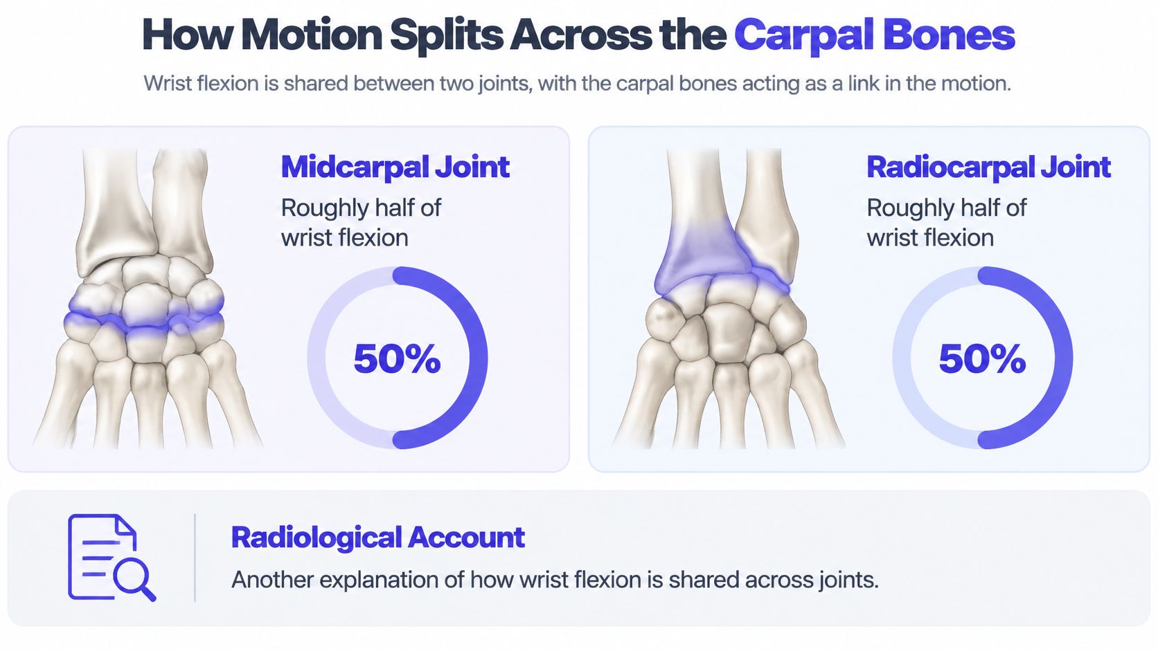
Wrist motion is shared across several joints, not produced by one hinge. The radiocarpal and midcarpal joints contribute to the arc together, while the carpal bones adjust their positions as the hand moves.
Approximately half of wrist flexion occurs at the midcarpal joint and the other half at the radiocarpal joint, according to this clinical anatomy text on wrist kinematics (opens in a new tab). That division describes the combined movement of the wrist, rather than a single bone rotating around a fixed axle.
Why the numbers differ between sources
The measured contribution of each joint can depend on how motion is recorded and where researchers draw the boundary between the two layers. Some methods track bone positions in planar images; others use in vivo imaging with the hand loaded. A figure is most useful when you know which joint layer it describes and how the measurement was made.
The carpal rows also differ in how they move. In vivo imaging shows that the distal carpal row behaves relatively rigidly, with less than 3 degrees of intercarpal motion during certain oblique wrist paths, as reported in this study of carpal kinematics under in vivo imaging (opens in a new tab). The proximal row, by contrast, adjusts its position between the radius and the distal row. Each carpal bone can translate and rotate, so the wrist's movement comes from coordinated changes across the bones.
Clinical consequence: A stiff or unstable region can alter how load passes through the wrist without removing its total range of motion. A wrist that bends through a full arc may still have altered movement within its carpal rows.
A goniometer records the hand's overall flexion arc. It does not show how much each joint layer contributed, or whether an individual carpal bone moved differently from its neighbours.
What Joint Type Does and Does Not Tell You About Wrist Pain
Classification predicts movement capacity. It does not predict the source of pain. These are separate questions, and conflating them is the second most common error students make with the wrist.
A 2025 review of MRI findings in wrist pain describes overlapping causes, including the triangular fibrocartilage complex, interosseous ligaments, ulnocarpal impaction and midcarpal instability, as summarised in the review of MRI in wrist pathology (opens in a new tab). Pain can arise from soft tissue or from several articulations interacting, not from the radiocarpal joint alone. The same review notes that radial-sided wrist pain is difficult to localise because several structures overlap, with MRI acting as an adjunct when examination findings are subtle or inconclusive.
Two misconceptions worth correcting
Misconception one: an MRI is automatically needed. It isn't. Plain radiographs are generally the initial test for acute wrist pain. MRI or ultrasound becomes appropriate when tendon or ligament injury is suspected, and CT is especially useful for fracture detail or alignment. Reaching for the most advanced scan first doesn't produce a better answer, it produces a slower one.
Misconception two: normal X-rays rule out serious injury. They don't. Plenty of ligament and tendon injuries look unremarkable on a plain film, which is why a normal X-ray plus ongoing pain is a reason for further assessment, not a reason to stop looking.
What narrows the question is the clinical picture. Where the pain sits matters: thumb-side pain near the scaphoid and radial styloid behaves differently from little-finger-side pain near the disc and ulnocarpal region. How it started matters, with a fall onto an outstretched hand pointing one way and a repetitive gripping load pointing another. Swelling, instability and which movements reproduce the pain all carry more diagnostic weight than the joint's classification.
A worked case makes it concrete. Say a student falls on an outstretched hand and reports pain only at the base of the thumb, with full range of motion and no swelling. The classification "ellipsoid joint" says nothing useful here. The location, the mechanism and the scaphoid's anatomy say a great deal, and the case study of hand pain in a 16-year-old shows how that reasoning is written up.
Bring three things to an appointment: the exact site you can point to with one finger, the mechanism of injury, and when the pain started.
That's more useful than any description of joint type you could offer.
Confirm the Joint Before You Answer
Before a label goes onto a diagram or into an exam answer, run this check in order.
- Scale. Does the question mean the radiocarpal joint or the whole wrist complex? Answer the scale you were asked about, and say which one you're using.
- Direction. Is the movement happening at the hand (flexion, extension, radial and ulnar deviation) or at the forearm (pronation, supination, which belong to the distal radioulnar joint)?
- Wording. Which classification term does your module use, and does its glossary match the textbook's? Course glossaries differ, and the module's own notes decide the mark.
- Completeness of the diagram. Are both carpal rows labelled? A diagram showing only the proximal row has left out the midcarpal joint.
An answer template for exams
Name the joint, state its classification, list its main movements, then add the exception that shows you understand the boundary. For example: the radiocarpal joint is a synovial ellipsoid (condyloid) joint permitting flexion, extension, radial and ulnar deviation; the midcarpal joint is classified the same way and contributes through intercarpal gliding; the thumb carpometacarpal joint is a saddle joint and is not part of the wrist complex proper.
That structure earns marks because it answers the question that was asked and pre-empts the follow-up.
The concrete next step is a joint-by-joint table in your own handwriting, one row per joint, four columns: articulation, type, movements, and one thing the joint does not do. Check it against past-paper answers and your university's course-specific notes for the module, and adjust the wording until it matches how your course defines each term.
If you want to check your joint-by-joint table against how your own module phrases things, CramShare holds course-matched anatomy notes, summaries and past-paper material organised by university, course code and textbook, so you can compare wording rather than guess at it. The CramShare library also lets you preview documents before you commit, which matters when two courses describe the same wrist differently. Build the table once, verify it against your module's terminology, and you'll answer wrist joint type questions without hesitating again.