Neonatal And Pediatric Respiratory Care, 5th Edition Test Bank

Preview Extract
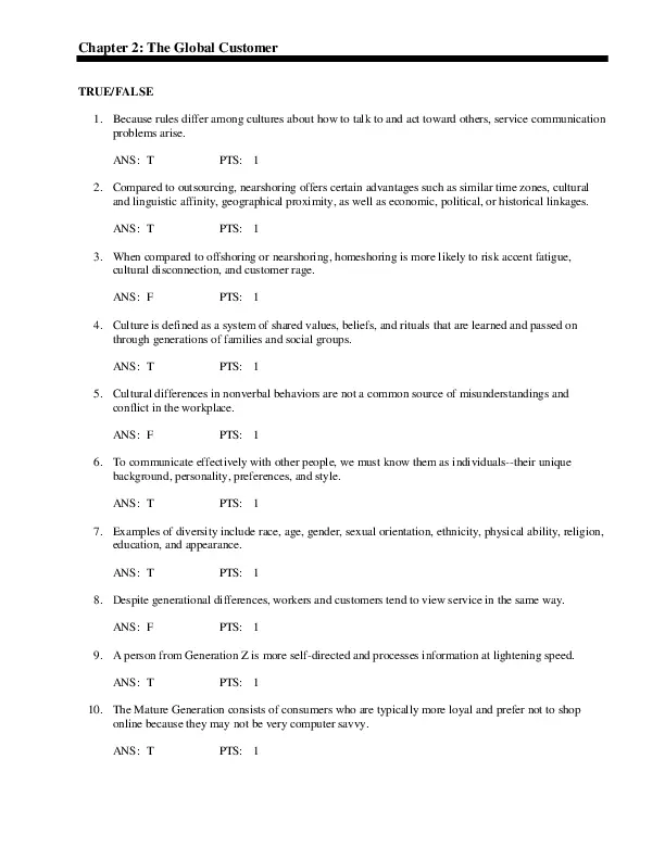
Chapter 02: Fetal Gas Exchange and Circulation Walsh: Neonatal and Pediatric Respiratory Care, 5th Edition MULTIPLE CHOICE 1. Which of the following embryonic germ layers gives formation to the respiratory system? a. Endoderm b. Mesoderm c. Ectoderm d. Periderm ANS: A The respiratory systemโ€”pharynx, lungs, and epithelial lining of the trachea and lungsโ€”originates in the endoderm. Refer to Box 2-1 in the textbook to see the list of various tissue systems found in the three embryonic layers. 2. What is the function of Whartonโ€™s jelly inside the umbilical cord? a. To help provide nutrition to the fetus b. To prevent the vessels inside the cord from kinking c. To help protect the fetus d. To regulate the temperature between the fetus and the mother ANS: B Whartonโ€™s jelly, a gelatinous substance inside the umbilical cord, helps protects the vessels of the fetus and may prevent the cord from kinking. 3. Which of the following organs is considered to be the first complete form? a. Heart b. Brain c. Lungs d. Kidneys ANS: A The heart is considered to be the first complete organ formed. By 8 weeks of gestation, the normal fetal heart is fully functional, complete with all chambers, valves, and major vessels. 4. A pregnant woman is coming for an early prenatal evaluation and wants to know if she can listen to the babyโ€™s heartbeat. How early can the fetal heartbeat be detected? a. Day 8 b. Day 22 c. Day 45 d. Day 60 ANS: B By day 22, cardiac contractions are detectable and bidirectional tidal blood flow begins. 5. Which of the following anatomic structures are fetal shunts? I. Foramen ovale II. Sinus venosus III. Ductus venosus IV. Ductus arteriosus a. I and III only b. I, III, and IV only c. I, II, and IV only d. II, III, and IV only ANS: B Figure 2-6 in the textbook illustrates fetal circulation and the three shunts present in the fetus that close soon after birth. They include (1) the foramen ovale, the opening between the right atrium and the left atrium, which enables oxygenated blood to flow to the left side of the fetal heart; (2) the ductus venosus, which appears continuous with the umbilical vein and shunts 30% to 50% of oxygen-rich blood around the liver; and (3) the ductus arteriosus, which allows most of the pulmonary arterial blood flow to bypass the nonfunctioning fetal lungs and enter the aorta. 6. Which of the following events causes cessation of right-to-left shunt through the foramen ovale? a. Increased levels of PO2 in the blood of the neonate b. Decreased levels of PCO2 in the blood of the newborn c. Increased systemic vascular resistance d. Removal of the placenta, causing lowered blood volume returning to the right side of the fetal heart ANS: C Once the cord is clamped and the PVR decreases, pressures in the right side of the heart decrease and pressures in the left side increase. Because the foramen ovale flap allows blood to flow only from right to left, it closes when the pressures in the left atrium become greater than those in the right atrium. Closing the foramen ovale further facilitates the increase of blood flow to the lungs during the transitional period and is necessary to maintain normal extrauterine circulation. 7. How long after birth should it take for the ductus arteriosus to close completely? a. 24 hours b. 48 hours c. 96 hours d. 1 week ANS: C Because the pressure in the aorta also increases and becomes greater than the pressure in the pulmonary artery, the amount of shunting through the ductus arteriosus decreases. The functional closure of the ductus arteriosus occurs as a result of being exposed to an increased PO2, a decrease in PVR leading to the reduction in blood pressure within the ductal lumen, a decrease in the local production of prostaglandins, and a reduction in the number of prostaglandin receptors within the tissue of the ductus arteriosus. Normally, constriction of the ductus arteriosus starts to occur at birth, and 20% of the ductus closes within 24 hours, with 80% closed in 48 hours, and 100% by 96 hours after birth. 8. During early embryonic development, which of the following structures supply nutrition to the growing embryo? a. Angiogenic clusters b. Chorionic membrane c. Bulbus cordis d. Whartonโ€™s jelly ANS: A During early embryonic development, small cellular pools, referred to as angiogenic clusters or blood islands, supply nutrition to the growing embryo. These clusters coalesce to form two heart tubes lined with specialized myocardial tissue. 9. Which of the following paths accurately traces oxygenated blood from the placenta to the fetus? a. Umbilical arteries, ductus venosus, right atrium, left atrium, foramen ovale, aorta, umbilical vein b. Umbilical vein, ductus arteriosus, right atrium, umbilical arteries, foramen ovale, left atrium, aorta c. Aorta, umbilical arteries, umbilical vein, ductus venosus, right atrium, foramen ovale, left atrium d. Umbilical vein, ductus venosus, right atrium, foramen ovale, left atrium, aorta, umbilical arteries ANS: D The path of fetal circulation and percentage of oxygen saturation in various locations is illustrated in Figure 2-6.

Document Preview (3 of 238 Pages)

User generated content is uploaded by users for the purposes of learning and should be used following SchloarOn's honor code & terms of service.
You are viewing preview pages of the document. Purchase to get full access instantly.

Shop by Category See All


Shopping Cart (0)

Your bag is empty

Don't miss out on great deals! Start shopping or Sign in to view products added.

Shop What's New Sign in