Comprehensive Radiographic Pathology 5th Edition Test Bank

Preview Extract
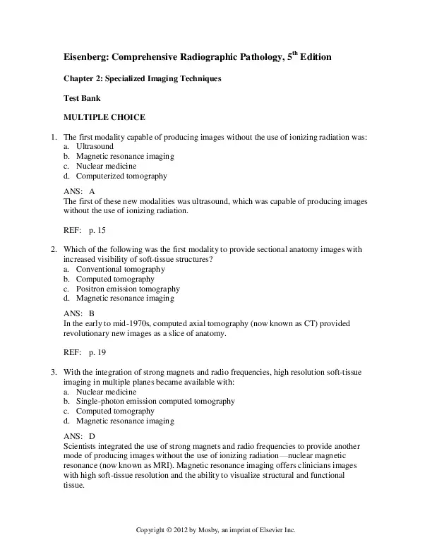
Eisenberg: Comprehensive Radiographic Pathology, 5th Edition Chapter 2: Specialized Imaging Techniques Test Bank MULTIPLE CHOICE 1. The first modality capable of producing images without the use of ionizing radiation was: a. Ultrasound b. Magnetic resonance imaging c. Nuclear medicine d. Computerized tomography ANS: A The first of these new modalities was ultrasound, which was capable of producing images without the use of ionizing radiation. REF: p. 15 2. Which of the following was the first modality to provide sectional anatomy images with increased visibility of soft-tissue structures? a. Conventional tomography b. Computed tomography c. Positron emission tomography d. Magnetic resonance imaging ANS: B In the early to mid-1970s, computed axial tomography (now known as CT) provided revolutionary new images as a slice of anatomy. REF: p. 19 3. With the integration of strong magnets and radio frequencies, high resolution soft-tissue imaging in multiple planes became available with: a. Nuclear medicine b. Single-photon emission computed tomography c. Computed tomography d. Magnetic resonance imaging ANS: D Scientists integrated the use of strong magnets and radio frequencies to provide another mode of producing images without the use of ionizing radiationโ€”nuclear magnetic resonance (now known as MRI). Magnetic resonance imaging offers clinicians images with high soft-tissue resolution and the ability to visualize structural and functional tissue. Copyright ยฉ 2012 by Mosby, an imprint of Elsevier Inc. Test Bank 2-2 REF: p. 15 4. Single-photon emission computed tomography took nuclear medicine imaging concepts and added the use of: a. New pharmaceuticals b. New pulsing sequences c. Gamma camera movement d. Stronger frequency transducers ANS: C Nuclear medicine expanded its role by adding movement and a computer that allowed more than anterior and posterior projections, resulting in the development of single-photon emission computed tomography (SPECT). REF: p. 16 5. Which of the following uses gamma-emitting radionuclides to produce images? a. X-ray images b. Nuclear medicine images c. PET images d. Sonography images ANS: B Gamma-emitting radionuclides are detected by the gamma camera to produce an image. REF: p. 27 6. Molecular imaging advancements using a positron-emitting radiopharmaceutical is: a. SPECT b. CAT c. MRI d. PET ANS: D The creation of a positron-emitting radionuclide resulted in the newest modalityโ€”positron emission tomography (PET). REF: p. 16 7. The superimposition of images from two different digital modalities produces images: a. Of anatomic and physiologic perspectives b. Using software for image integration c. From hybrid equipment, such as PET-CT d. All of the above ANS: D Copyright ยฉ 2012 by Mosby, an imprint of Elsevier Inc. Test Bank 2-3 Imaging modalities with special software can now be integrated to create a fused image (superimposition of two different modalities). PET-CT is the most prominent hybrid equipment available today. REF: p. 16 8. The complexity of technology requires the radiographer to: a. Have a more specific skill set b. Have an extensive knowledge of anatomy c. Have computer networking classes d. A and B ANS: D Complex technology requires the radiographer to have a broader and more specific skill set to produce quality images. REF: p. 15 9. Which of the following specialized imaging techniques provides cross-sectional images at a low cost, is readily available, and has the ability to differentiate cystic, solid, and complex tissue? a. Sonography b. CT c. MRI d. PET ANS: A Ultrasound (sonography) is a widely accepted cross-sectional imaging technique because of its low cost, availability, and ability to differentiate cystic (gallbladder), solid (liver), and complex (liver tumor) tissue. REF: p. 16 10. In ultrasound the high frequency sound waves are produced by: a. Sound waves emanating from the body b. Electrical stimulation of a specialized crystal c. Magnets and pulse radiofrequencies d. Radiopharmaceuticals ANS: B Ultrasound relies on high-frequency sound waves produced by electrical stimulation of a specialized crystal to produce an image. REF: p. 16 11. Reflections in ultrasound produced by tissue interaction are called: a. Isoechoic b. Echoes Copyright ยฉ 2012 by Mosby, an imprint of Elsevier Inc. Test Bank 2-4 c. Pulses d. Frequencies ANS: B The transducer records the tiny changes of the signalโ€™s pitch and direction. A water-tissue interface can produce strong reflections (echoes), whereas a solid tissue mass that contains small differences in composition can cause weak reflections. REF: p. 16 12. Fluid-filled structures transmitting sound waves easily and appearing as a dark region on the image are _____ tissue. a. Hyperechoic b. Hypoechoic c. Anechoic d. Isoechoic ANS: C Anechoic tissue or structures (which are echo free or lacking a signal) transmit sound waves easily and appear as the dark region on the image. REF: p. 17 13. The major advantage of ultrasound is: a. No adverse effects on human tissue b. Ability to image anatomy in cross-section c. Ionizing radiation is used to produce an image d. Only low energy gamma radiation is used to produce an image ANS: A The major advantage of ultrasound is its safety. REF: p. 17 14. To produce quantitative data in ultrasound, the equipment must: a. Integrate real-time imaging with sono CT b. Include harmonic technology c. Fuse conventional real-time with Doppler imaging d. Have a multifrequency transducer ANS: C The color-flow duplex system, in which conventional real-time imaging is integrated with Doppler imaging (to produce quantitative data) REF: p. 18 15. To demonstrate stenoses, blood clots, plaques, and emboli requires: a. A multifrequency transducer Copyright ยฉ 2012 by Mosby, an imprint of Elsevier Inc. Test Bank 2-5 b. A color-flow duplex system c. Sono CT real-time compound imaging d. Harmonic imaging ANS: B Vascular or color-flow Doppler studies assess the patency of major blood vessels, demonstrating obstructions (stenoses), blood clots, plaques, and emboli REF: p. 18 16. Using a narrow x-ray beam and scanning from multiple angles, the amount of radiation absorbed in tissue is represented by: a. A relative linear attenuation coefficient b. Data for computer reconstruction c. A gray scale on the computer monitor d. All of the above ANS: D Scanning a slice of tissue from multiple angles with a narrow x-ray beam, then calculating a relative linear attenuation coefficient (representing the amount of radiation absorbed in tissue for the various tissue elements in the section), and finally displaying the computed reconstruction as a gray-scale image on a television monitor is CT. REF: p. 19 17. The attenuation of a specific tissue relative to water is the: a. CT number b. Echo coefficient c. Positron absorption d. Pulse sequence ANS: A The CT number reflects the attenuation of a specific tissue relative to that of water, which is arbitrarily assigned a CT number of 0 and appears gray on the image. REF: p. 19 18. Window width in CT controls: a. Density b. Gray scale c. Midrange of densities d. Resolution ANS: B Window widths control gray scaleโ€”contrast scale and window levels control density. From the radiographerโ€™s perspective, this means the window width determines the number of densities that will be visualized on the monitor. Copyright ยฉ 2012 by Mosby, an imprint of Elsevier Inc. Test Bank 2-6 REF: p. 19 19. The midpoint or center of the number of densities being viewed in a selected window on CT is the: a. Window width b. Pulse width c. Window level d. X-ray level ANS: C The window level is the midpoint or center of the total number of densities being viewed in a selected window width. REF: p. 19 20. High-resolution CT requires the slice sections to be: a. 1.5 to 2.0 mm b. 2 to 5 mm c. 5 to 10 mm d. 1 to 2 cm ANS: A In high-resolution CT, thin sections (1.5 to 2.0 mm slices) are used to produce a very detailed display of lung anatomy. REF: p. 20 21. CT scanning permitting data that can easily be reformatted to produce multiplanes (coronal and sagittal) is accomplished by: a. High-resolution scans b. Conventional scans c. Helical scanning d. Single-scan protocols ANS: C Spiral (helical) scanning is the technique where continual CT scanning is performed as the patient moves through the gantry (unlike the multiple single scans in conventional CT). This permits much faster scanning without respiratory motion and provides data that can be easily reformatted in coronal and sagittal planes. REF: p. 20 22. What made CT angiography more precise and prevalent? a. The development of 16-slice (and greater) scanners b. Multiplanar images produced by multifrequency transducers c. Three-dimensional images d. A and C Copyright ยฉ 2012 by Mosby, an imprint of Elsevier Inc. Test Bank 2-7 ANS: D Using volume-rendered imaging and 3-D volume rendering, the vascular system can be viewed from all perspectives (360ยฐ). CT angiography is more prevalent and precise as a result of the development of 16-slice and greater scanners. REF: p. 21 23. All of the following are post-processing techniques used in CT, except: a. Maximum intensity projections b. Shaded surface rendering c. Time-of-flight technique d. Volume-rendering ANS: C Time-of-flight is an angiographic technique used with MRI. REF: pp. 22-23 24. The modality of choice to image the central nervous system is: a. CT b. MRI c. Ultrasound d. PET ANS: B MRI has become the modality of choice for imaging the central nervous system and spine. REF: p. 23 25. What atomโ€™s energy is manipulated to produce MRI images? a. radiofrequency pulse (RF) hydrogen b. Echo time (TE) carbon c. Relaxation time (T1/T2) nitrogen d. Repetition time (TR) oxygen ANS: A The basic technique consists of inducing hydrogen atoms (protons) to alternate between a high-energy state and a low-energy state by absorbing and then releasing, or transferring, energy. This absorption of energy is accomplished by placing the anatomic part to be imaged in a strong static magnetic field and directing a radiofrequency (RF) pulse at the area REF: p. 23 26. Relaxation is: a. The energy transfer of the radiofrequency pulse to tissue b. The proton releasing energy to return to its original state Copyright ยฉ 2012 by Mosby, an imprint of Elsevier Inc. Test Bank 2-8 c. The listening time of the MR unit to hear an echo d. The repetition of the energy signal ANS: B The protons begin to release, or transfer, their absorbed energy as they move back to a low-energy state. This process is called relaxation, and it occurs over time. REF: p. 23 27. By selecting different pulses, sequences, and frequencies, the MR T1-weighted image produces images with a: a. High signal intensity (bright) water b. High signal intensity (bright) fat, subacute hemorrhage, and contrast material c. Low signal intensity (intermediate to dark) fat d. Low signal intensity (very dark) bone and air ANS: B On T1-weighted images, substances causing high signal intensity (that is, appearing bright) include fat, subacute hemorrhage, highly proteinaceous material (for example, . mucus), slow-flowing blood, and intravenous contrast material (for example,.g, gadolinium). REF: pp. 23-24 28. To best increase the exam specificity in MRI, the technologist must: a. Use contrast enhancement b. Change relaxation time c. Change pulse sequences d. A and C ANS: D The introduction of intravenous contrast materials and different types of pulse sequences are helping increase specificity. REF: p. 24 29. Diffusion imaging relies on _____ aiding in diagnosis of early stroke. a. Tissue diffused with contrast material b. Decreased microcirculation demonstrating less blood volume c. Random movement of water d. A and C ANS: C Diffusion imaging relies on the movement of molecules and random thermal motion. (In this instance, random movement of water is known as diffusion.) REF: p. 26 Copyright ยฉ 2012 by Mosby, an imprint of Elsevier Inc. Test Bank 2-9 30. Motor, sensory, and vision neurons (nervous system) can be demonstrated by: a. Diffusion imaging b. Perfusion imaging c. Fat-suppressed images d. Functional MR ANS: D Functional MR (fMR) allows the localization of specific regions of the brain that correspond to various functions, such as the motor, sensory, memory, vision, and language functions. REF: p. 26 31. Ionizing radiation, which is emitted from the patient to produce images, is the modality of: a. Conventional x-ray b. Ultrasound c. Nuclear medicine d. Magnetic resonance imaging ANS: C In nuclear medicine, the patient ingests, or is injected with, a radiopharmaceutical that emits radiation, and an image is created from the signals radiating from the patient. REF: p. 27 32. The patient is injected or ingests the ionizing radiation through: a. A radiopharmaceutical b. An iodinated contrast agent c. Gadolinium d. Barium ANS: A The dose of radiopharmaceutical is calculated on the basis of the specific half-life and decay rate of its attached radionuclide. The amount of ionizing radiation to the patient in a nuclear medicine study is similar to that in a plain radiographic examination. REF: p. 27 33. To detect the ionizing radiation being emitted from the patient to produce an image, a _____ is used. a. Geiger counter b. Gamma camera c. Pocket dosimeter d. Dose calibrator ANS: B Copyright ยฉ 2012 by Mosby, an imprint of Elsevier Inc. Test Bank 2-10 A gamma camera with a sodium iodide crystal detects the ionizing radiation emitted from the patient. REF: p. 27 34. To best demonstrate a physiologic map, the modality of choice is: a. MRI b. CT c. Ultrasound d. Nuclear medicine ANS: D The physiologic map produced by some nuclear medicine procedures allows changes to be detected earlier than plain radiographic images because the functional perspective makes it more sensitive. REF: p. 28 35. Increased radionuclide uptake produces a _____ on the image. a. Hot spot b. Blank spot c. Cold spot d. Homogeneous area ANS: A Abnormal nuclear medicine images demonstrate hot spots produced by an increase in the uptake. REF: p. 28 36. To enhance the nuclear medicine image, researchers applied the use of _____ to increase accuracy. a. Computers and movement b. Multiple gamma cameras and movement c. Multiple digital cameras d. A and B ANS: D SPECT imaging involves the movement of the gamma camera(s) rotating around the patient and the computer software to perform the reconstruction of the image. REF: pp. 28-29 37. A collimator in the SPECT imaging system: a. Is multiple parallel channels allowing rays to pass b. Provides a lead plate increasing image contrast c. Is a device to allow rotation of the camera d. Allows more gamma rays to reach the camera Copyright ยฉ 2012 by Mosby, an imprint of Elsevier Inc. Test Bank 2-11 ANS: A SPECT has a collimator containing multiple parallel channels to allow the rays to pass. REF: p. 29 38. The greatest difference in PET and SPECT is the type of: a. Ionizing radiation emitted by radiopharmaceuticals b. Camera used to detect the ionizing radiation c. Physiologic map produced d. Computer images produced ANS: A In positron emission tomography (PET), as in nuclear medicine procedures, a radionuclide tracer is used to produce images. However, here the radiopharmaceutical is different because it decays by positron emission. REF: p. 29 39. Epileptic foci and Alzheimerโ€™s disease is best demonstrated by: a. SPECT b. CT c. MRI d. PET ANS: D PET can be used to evaluate for stroke and to identify epileptic foci for surgical intervention. Brain disorders that can be demonstrated by PET imaging include Parkinsonโ€™s disease, schizophrenia, Huntingtonโ€™s disease, and Alzheimerโ€™s disease. REF: p. 31 40. Which imaging technique analyzes the chemical composition of tissues in vivo? a. CTA b. MRA c. fMRI d. MR spectroscopy ANS: D The development of magnetic resonance spectroscopy (MRS) has made is possible to analyze the chemical composition of tissues in vivo. REF: p. 25 MATCHING Match the following terms related to specialty imaging technologies. a. Gamma camera movement and computer enhancement Copyright ยฉ 2012 by Mosby, an imprint of Elsevier Inc. Test Bank b. c. d. e. f. g. h. 1. 2. 3. 4. 5. 6. 7. 8. 2-12 Hybrid equipment producing two modality images simultaneously Modality of choice for examinations of children and pregnant women Radiofrequency pulses used to excite protons in the atoms Radiopharmaceutical emitting gamma radiation to produce an image Scanning a slice of tissue from multiple angles with a narrow x-ray beam Software specially designed to overlay multidimensional computed data Uses a decaying radiopharmaceutical emitting a positron Computed tomography Direct fusion imaging Integrated imaging Magnetic resonance imaging Nuclear medicine PET SPECT Ultrasound 1. ANS: F REF: p. 19 NOT: Rationale: Scanning a slice of tissue from multiple angles with a narrow x-ray beam, then calculating a relative linear attenuation coefficient (representing the amount of radiation absorbed in tissue for the various tissue elements in the section), and finally displaying the computed reconstruction as a gray-scale image on a television monitor is CT. Hybrid technology-two modalities scanning simultaneously-produces a direct fusion image. Integrated images are a fusion of multidimensional data from MR, CT, NM, SPECT, or PET to create a single set of images using software. The basic technique of MRI consists of inducing hydrogen atoms (protons) to alternate between a high-energy state and a low-energy state by absorbing and then releasing, or transferring, energy. Gamma-emitting radionuclides are detected by the gamma camera to produce an image. 2. ANS: B REF: p. 32 NOT: Rationale: Scanning a slice of tissue from multiple angles with a narrow x-ray beam, then calculating a relative linear attenuation coefficient (representing the amount of radiation absorbed in tissue for the various tissue elements in the section), and finally displaying the computed reconstruction as a gray-scale image on a television monitor is CT. Hybrid technology-two modalities scanning simultaneously-produces a direct fusion image. Integrated images are a fusion of multidimensional data from MR, CT, NM, SPECT, or PET to create a single set of images using software. The basic technique of MRI consists of inducing hydrogen atoms (protons) to alternate between a high-energy state and a low-energy state by absorbing and then releasing, or transferring, energy. Gamma-emitting radionuclides are detected by the gamma camera to produce an image. 3. ANS: G REF: p. 32 NOT: Rationale: Scanning a slice of tissue from multiple angles with a narrow x-ray beam, then calculating a relative linear attenuation coefficient (representing the amount of radiation absorbed in tissue for the various tissue elements in the section), and finally displaying the computed reconstruction as a gray-scale image on a television monitor is CT. Hybrid technology-two modalities scanning simultaneously-produces a direct fusion image. Integrated images are a fusion of multidimensional data from MR, CT, NM, Copyright ยฉ 2012 by Mosby, an imprint of Elsevier Inc. Test Bank 4. 5. 6. 7. 2-13 SPECT, or PET to create a single set of images using software. The basic technique of MRI consists of inducing hydrogen atoms (protons) to alternate between a high-energy state and a low-energy state by absorbing and then releasing, or transferring, energy. Gamma-emitting radionuclides are detected by the gamma camera to produce an image. ANS: D REF: p. 23 NOT: Rationale: Scanning a slice of tissue from multiple angles with a narrow x-ray beam, then calculating a relative linear attenuation coefficient (representing the amount of radiation absorbed in tissue for the various tissue elements in the section), and finally displaying the computed reconstruction as a gray-scale image on a television monitor is CT. Hybrid technology-two modalities scanning simultaneously-produces a direct fusion image. Integrated images are a fusion of multidimensional data from MR, CT, NM, SPECT, or PET to create a single set of images using software. The basic technique of MRI consists of inducing hydrogen atoms (protons) to alternate between a high-energy state and a low-energy state by absorbing and then releasing, or transferring, energy. Gamma-emitting radionuclides are detected by the gamma camera to produce an image. ANS: E REF: pp. 27-28 NOT: Rationale: Scanning a slice of tissue from multiple angles with a narrow x-ray beam, then calculating a relative linear attenuation coefficient (representing the amount of radiation absorbed in tissue for the various tissue elements in the section), and finally displaying the computed reconstruction as a gray-scale image on a television monitor is CT. Hybrid technology-two modalities scanning simultaneously-produces a direct fusion image. Integrated images are a fusion of multidimensional data from MR, CT, NM, SPECT, or PET to create a single set of images using software. The basic technique of MRI consists of inducing hydrogen atoms (protons) to alternate between a high-energy state and a low-energy state by absorbing and then releasing, or transferring, energy. Gamma-emitting radionuclides are detected by the gamma camera to produce an image. ANS: H REF: pp. 29-30 NOT: Rationale: Scanning a slice of tissue from multiple angles with a narrow x-ray beam, then calculating a relative linear attenuation coefficient (representing the amount of radiation absorbed in tissue for the various tissue elements in the section), and finally displaying the computed reconstruction as a gray-scale image on a television monitor is CT. Hybrid technology-two modalities scanning simultaneously-produces a direct fusion image. Integrated images are a fusion of multidimensional data from MR, CT, NM, SPECT, or PET to create a single set of images using software. The basic technique of MRI consists of inducing hydrogen atoms (protons) to alternate between a high-energy state and a low-energy state by absorbing and then releasing, or transferring, energy. Gamma-emitting radionuclides are detected by the gamma camera to produce an image. ANS: A REF: pp. 28-29 NOT: Rationale: Scanning a slice of tissue from multiple angles with a narrow x-ray beam, then calculating a relative linear attenuation coefficient (representing the amount of radiation absorbed in tissue for the various tissue elements in the section), and finally displaying the computed reconstruction as a gray-scale image on a television monitor is CT. Hybrid technology-two modalities scanning simultaneously-produces a direct fusion image. Integrated images are a fusion of multidimensional data from MR, CT, NM, SPECT, or PET to create a single set of images using software. The basic technique of MRI consists of inducing hydrogen atoms (protons) to alternate between a high-energy Copyright ยฉ 2012 by Mosby, an imprint of Elsevier Inc. Test Bank 2-14 state and a low-energy state by absorbing and then releasing, or transferring, energy. Gamma-emitting radionuclides are detected by the gamma camera to produce an image. 8. ANS: C REF: p. 17 NOT: Rationale: Scanning a slice of tissue from multiple angles with a narrow x-ray beam, then calculating a relative linear attenuation coefficient (representing the amount of radiation absorbed in tissue for the various tissue elements in the section), and finally displaying the computed reconstruction as a gray-scale image on a television monitor is CT. Hybrid technology-two modalities scanning simultaneously-produces a direct fusion image. Integrated images are a fusion of multidimensional data from MR, CT, NM, SPECT, or PET to create a single set of images using software. The basic technique of MRI consists of inducing hydrogen atoms (protons) to alternate between a high-energy state and a low-energy state by absorbing and then releasing, or transferring, energy. Gamma-emitting radionuclides are detected by the gamma camera to produce an image. Match the following terms related to ultrasound. a. Echo free appearing as a dark region on the image b. Reflections from body structures c. Structure exhibits a relatively strong reflection appearing light gray to white d. Tissue producing weak reflections that appear gray on the image e. Two structures producing the same reflection 9. 10. 11. 12. 13. Anechoic Echoes Echogenic Hypoechoic Isoechoic 9. ANS: A REF: pp. 16-17 NOT: Rationale: Anechoic is echo free or lacking a signal. Echoes are produced by water-tissue interface resulting in a strong reflection. Echogenic indicates a tissue structure producing a relatively strong reflection. Hypoechoic indicates a tissue structure producing a relatively weak reflection. Isoechoic reflections are two structures with the same echogenicity. 10. ANS: B REF: pp. 16-17 NOT: Rationale: Anechoic is echo free or lacking a signal. Echoes are produced by water-tissue interface resulting in a strong reflection. Echogenic indicates a tissue structure producing a relatively strong reflection. Hypoechoic indicates a tissue structure producing a relatively weak reflection. Isoechoic reflections are two structures with the same echogenicity. 11. ANS: C REF: pp. 16-17 NOT: Rationale: Anechoic is echo free or lacking a signal. Echoes are produced by water-tissue interface resulting in a strong reflection. Echogenic indicates a tissue structure producing a relatively strong reflection. Hypoechoic indicates a tissue structure producing a relatively weak reflection. Isoechoic reflections are two structures with the same echogenicity. 12. ANS: D REF: pp. 16-17 Copyright ยฉ 2012 by Mosby, an imprint of Elsevier Inc. Test Bank 2-15 NOT: Rationale: Anechoic is echo free or lacking a signal. Echoes are produced by water-tissue interface resulting in a strong reflection. Echogenic indicates a tissue structure producing a relatively strong reflection. Hypoechoic indicates a tissue structure producing a relatively weak reflection. Isoechoic reflections are two structures with the same echogenicity. 13. ANS: E REF: pp. 16-17 NOT: Rationale: Anechoic is echo free or lacking a signal. Echoes are produced by water-tissue interface resulting in a strong reflection. Echogenic indicates a tissue structure producing a relatively strong reflection. Hypoechoic indicates a tissue structure producing a relatively weak reflection. Isoechoic reflections are two structures with the same echogenicity. Match the following terms related to computed tomography. a. Ability to collect data from multiple sections per rotation b. Attenuation of a specific tissue relative to water c. Calculation of data collected from multiple angles using a narrow x-ray beam d. Continuous scanning while the patient moves through the gantry e. Contrast scale to be demonstrated on computer monitor f. Midpoint selected of densities to be viewed on the computer monitor g. Single scans that are integrated to produce multiplane images 14. 15. 16. 17. 18. 19. 20. Conventional scanning CT number Helical scanning Multidetection scanning Relative linear attenuation coefficient Window level Window width 14. ANS: G REF: p. 19 NOT: Rationale: Multidetection scanning collects data by multiple slices per rotation. The CT number reflects the attenuation of a specific tissue relative to that of water, which is arbitrarily assigned a CT number of 0 and appears gray on the image. Scanning a slice of tissue from multiple angles with a narrow x-ray beam, then calculating a relative linear attenuation coefficient (representing the amount of radiation absorbed in tissue for the various tissue elements in the section), and finally displaying the computed reconstruction as a gray-scale image on a television monitor is conventional CT. Spiral (helical) scanning is the technique where continual CT scanning is performed as the patient moves through the gantry (unlike the multiple single scans in conventional CT). This permits much faster scanning without respiratory motion and provides data that can be easily reformatted in coronal and sagittal planes. 15. ANS: B REF: p. 19 NOT: Rationale: Multidetection scanning collects data by multiple slices per rotation. The CT number reflects the attenuation of a specific tissue relative to that of water, which is arbitrarily assigned a CT number of 0 and appears gray on the image. Scanning a slice of tissue from multiple angles with a narrow x-ray beam, then calculating a relative linear Copyright ยฉ 2012 by Mosby, an imprint of Elsevier Inc. Test Bank 2-17 is arbitrarily assigned a CT number of 0 and appears gray on the image. Scanning a slice of tissue from multiple angles with a narrow x-ray beam, then calculating a relative linear attenuation coefficient (representing the amount of radiation absorbed in tissue for the various tissue elements in the section), and finally displaying the computed reconstruction as a gray-scale image on a television monitor is conventional CT. Spiral (helical) scanning is the technique where continual CT scanning is performed as the patient moves through the gantry (unlike the multiple single scans in conventional CT). This permits much faster scanning without respiratory motion and provides data that can be easily reformatted in coronal and sagittal planes. 20. ANS: E REF: p. 19 NOT: Rationale: Multidetection scanning collects data by multiple slices per rotation. The CT number reflects the attenuation of a specific tissue relative to that of water, which is arbitrarily assigned a CT number of 0 and appears gray on the image. Scanning a slice of tissue from multiple angles with a narrow x-ray beam, then calculating a relative linear attenuation coefficient (representing the amount of radiation absorbed in tissue for the various tissue elements in the section), and finally displaying the computed reconstruction as a gray-scale image on a television monitor is conventional CT. Spiral (helical) scanning is the technique where continual CT scanning is performed as the patient moves through the gantry (unlike the multiple single scans in conventional CT). This permits much faster scanning without respiratory motion and provides data that can be easily reformatted in coronal and sagittal planes. Match the following terms related to nuclear medicine. a. Annihilation produces two high-energy photons in opposite directions b. Distribution of radionuclide representing the organ or system c. Drug or chemical emitting radiation d. Interaction of gamma rays with the sodium iodide crystal to produce light e. Multiple gamma cameras with movement f. Sodium iodide crystal to detect ionizing radiation emitted from patient 21. 22. 23. 24. 25. 26. Gamma camera Physiologic map Positron emission tomography Radiopharmaceutical Scintigraphic image Single-photon emission computed tomography 21. ANS: F REF: p. 27 NOT: Rationale: Annihilation is the interaction producing two high-energy photons (gamma rays) in opposite directions (separated by 180ยฐ). In nuclear medicine, however, the patient ingests, or is injected with, a radiopharmaceutical that emits radiation, and an image is created from the signals radiating from the patient. A gamma camera with a sodium iodide crystal detects the ionizing radiation emitted from the patient. Interaction of the gamma rays with the crystal produces light scintillation, which is converted to a digital signal on a computer monitor. The scintigraphic image, which defines the distribution of the radioactive nuclide, represents the physiologic map of the organ or Copyright ยฉ 2012 by Mosby, an imprint of Elsevier Inc. Test Bank 22. 23. 24. 25. 2-18 system being imaged. SPECT imaging involves the movement of the gamma camera(s) rotating around the patient and the computer software to perform the reconstruction of the image. ANS: B REF: p. 28 NOT: Rationale: Annihilation is the interaction producing two high-energy photons (gamma rays) in opposite directions (separated by 180ยฐ). In nuclear medicine, however, the patient ingests, or is injected with, a radiopharmaceutical that emits radiation, and an image is created from the signals radiating from the patient. A gamma camera with a sodium iodide crystal detects the ionizing radiation emitted from the patient. Interaction of the gamma rays with the crystal produces light scintillation, which is converted to a digital signal on a computer monitor. The scintigraphic image, which defines the distribution of the radioactive nuclide, represents the physiologic map of the organ or system being imaged. SPECT imaging involves the movement of the gamma camera(s) rotating around the patient and the computer software to perform the reconstruction of the image. ANS: A REF: p. 29 NOT: Rationale: Annihilation is the interaction producing two high-energy photons (gamma rays) in opposite directions (separated by 180ยฐ). In nuclear medicine, however, the patient ingests, or is injected with, a radiopharmaceutical that emits radiation, and an image is created from the signals radiating from the patient. A gamma camera with a sodium iodide crystal detects the ionizing radiation emitted from the patient. Interaction of the gamma rays with the crystal produces light scintillation, which is converted to a digital signal on a computer monitor. The scintigraphic image, which defines the distribution of the radioactive nuclide, represents the physiologic map of the organ or system being imaged. SPECT imaging involves the movement of the gamma camera(s) rotating around the patient and the computer software to perform the reconstruction of the image. ANS: C REF: p. 16 | p. 27 NOT: Rationale: Annihilation is the interaction producing two high-energy photons (gamma rays) in opposite directions (separated by 180ยฐ). In nuclear medicine, however, the patient ingests, or is injected with, a radiopharmaceutical that emits radiation, and an image is created from the signals radiating from the patient. A gamma camera with a sodium iodide crystal detects the ionizing radiation emitted from the patient. Interaction of the gamma rays with the crystal produces light scintillation, which is converted to a digital signal on a computer monitor. The scintigraphic image, which defines the distribution of the radioactive nuclide, represents the physiologic map of the organ or system being imaged. SPECT imaging involves the movement of the gamma camera(s) rotating around the patient and the computer software to perform the reconstruction of the image. ANS: D REF: p. 28 NOT: Rationale: Annihilation is the interaction producing two high-energy photons (gamma rays) in opposite directions (separated by 180ยฐ). In nuclear medicine, however, the patient ingests, or is injected with, a radiopharmaceutical that emits radiation, and an image is created from the signals radiating from the patient. A gamma camera with a sodium iodide crystal detects the ionizing radiation emitted from the patient. Interaction of the gamma rays with the crystal produces light scintillation, which is converted to a Copyright ยฉ 2012 by Mosby, an imprint of Elsevier Inc. Test Bank 2-19 digital signal on a computer monitor. The scintigraphic image, which defines the distribution of the radioactive nuclide, represents the physiologic map of the organ or system being imaged. SPECT imaging involves the movement of the gamma camera(s) rotating around the patient and the computer software to perform the reconstruction of the image. 26. ANS: E REF: p. 16 | p. 28 NOT: Rationale: Annihilation is the interaction producing two high-energy photons (gamma rays) in opposite directions (separated by 180ยฐ). In nuclear medicine, however, the patient ingests, or is injected with, a radiopharmaceutical that emits radiation, and an image is created from the signals radiating from the patient. A gamma camera with a sodium iodide crystal detects the ionizing radiation emitted from the patient. Interaction of the gamma rays with the crystal produces light scintillation, which is converted to a digital signal on a computer monitor. The scintigraphic image, which defines the distribution of the radioactive nuclide, represents the physiologic map of the organ or system being imaged. SPECT imaging involves the movement of the gamma camera(s) rotating around the patient and the computer software to perform the reconstruction of the image. Copyright ยฉ 2012 by Mosby, an imprint of Elsevier Inc.

Document Preview (19 of 573 Pages)

User generated content is uploaded by users for the purposes of learning and should be used following SchloarOn's honor code & terms of service.
You are viewing preview pages of the document. Purchase to get full access instantly.

Shop by Category See All


Shopping Cart (0)

Your bag is empty

Don't miss out on great deals! Start shopping or Sign in to view products added.

Shop What's New Sign in